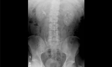
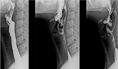
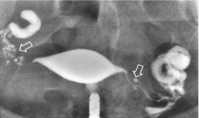
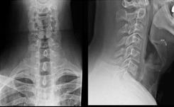
Digital X-ray and Procedures.

Diagnostic X-ray, or radiography, is a special method for taking pictures of areas inside the body. A machine focuses a small amount of radiation on the area of the body to be examined. The X-ray pass through the body, creating an image on film or a computer display.

The equipment, staff, and steps involved are different for each type of diagnostic X-ray procedure. However, they are all invaluable tools in detecting abnormalities and making early diagnosis of diseases or injury.

Key Benefits:

  • • Faster Results – Images are available instantly, reducing waiting time for diagnosis.
  • • Lower Radiation Exposure – Uses significantly less radiation compared to traditional X-rays.
  • • High Image Quality – Provides clearer and more detailed images for accurate diagnosis.
  • • Easy Image Storage & Sharing – Digital files can be stored, accessed, and shared quickly with specialists.
  • • Enhanced Diagnostic Accuracy – Advanced software allows zooming and image enhancement for better evaluation.
  • • Environment Friendly – Eliminates the need for films and chemicals used in conventional X-rays.
Whole Abdomen
IVP

IVP - An intravenous pyelogram (IVP), also called an intravenous urogram (IVU), is a radiological procedure used to observe the abnormalities of the urinary system, including the kidneys, ureters and bladder. Unlike X-ray KUB (Kidneys, Ureters and Bladder), which is a plain/noncontrast radiograph, an IVP is a diagnostic X-ray that brings contrast to highlight the urinary tract. In IVP after intravenous administration of non ionic contrast agent, radiographs obtained at specific intervals to visualise the urinary tract.

Pelvis
Barium Swallow

Barium swallow is an X-ray imaging test used to obtain a clear view of the structures of the esophagus. The patient swallows liquid barium while X-ray images are obtained. One can visualize the interior of the upper gastrointestinal tract using the radio-opaque substance barium. The barium fills and then coats the lining of the esophagus so that it can identify anatomical abnormalities such as tumors.

Upper Abdomen
HSG

A hysterosalpingogram or HSG is an X-ray procedure performed to assess whether the fallopian tubes are patent / open and take a look if the shape of the uterine cavity is normal An HSG is an outpatient procedure that will hardly take half an hour to perform rooutinely. Once your menstrual cycles have ended and before ovulation (usually 8-9 th day), this procedure is being performed to prevent interference with an early pregnancy.