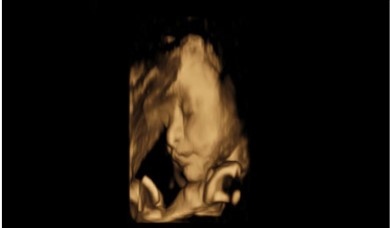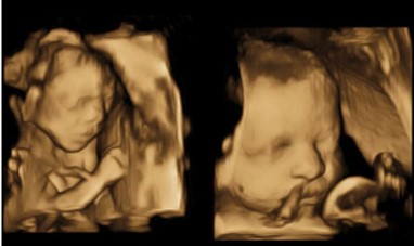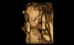
3D/4D Ultrasonography
3D/4D ultrasonography is an advanced imaging technique that provides three-dimensional and real-time (4D) images of the fetus during pregnancy or various organs in the body. Unlike traditional 2D ultrasounds, 3D imaging captures detailed, life-like still images, while 4D allows for live video, capturing movements in real time
This cutting-edge technology is particularly popular in prenatal care, offering expectant parents a glimpse of their unborn baby with stunning clarity. It's also used for more precise diagnosis of structural abnormalities in different parts of the body.
Key Benefits:
- • Bringing Life into Focus – See Tomorrow's Moments Today.
- • Real-Time Miracles – Experience the Future of Imaging
- • Discover Every Detail – 3D/4D Imaging for Clearer Answers