Echocardiography
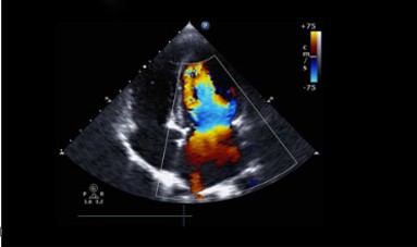
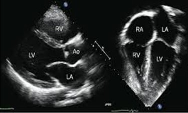
Echocardiography is a non-invasive diagnostic test that uses ultrasound waves to create detailed images of the heart's structure and function. It helps in assessing heart conditions by visualizing the heart chambers, valves, and blood flow.
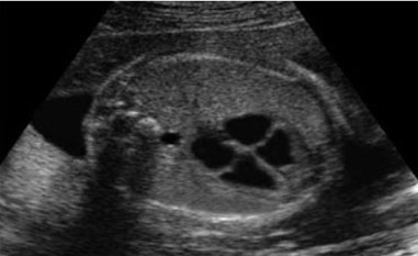
Commonly used to diagnose conditions such as heart valve problems, heart failure, congenital heart defects, and cardiomyopathies, echocardiography is a vital tool in cardiology
Key Benefits:
- • Non-invasive & Safey – Uses ultrasound waves with no radiation, making it completely safe.
- • Real-time Heart Imaging – rovides live images of the heart’s structure and function.
- • Detects Heart Conditions Early – Helps identify valve defects, heart failure, and congenital abnormalities.
- • Evaluates Blood Flow – Assesses blood circulation through the heart using Doppler imaging.
- • Guides Treatment Decisions – Assists doctors in planning and monitoring cardiac treatments.
- • Quick & Painless Procedure –Fast, comfortable, and requires no special recovery time.